
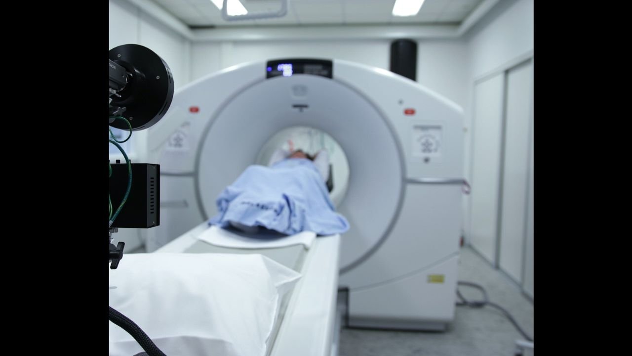
A new study published in The Journal of Nuclear Medicine in July 2026 has highlighted the potential value of repeating a prostate-specific membrane antigen (PSMA) PET scan in selected prostate cancer patients. Researchers found that a second scan identified previously undetected cancer in more than half of patients whose initial PSMA PET scan showed no evidence of disease.
The findings could influence how doctors manage patients experiencing rising prostate-specific antigen (PSA) levels after treatment, particularly when earlier imaging fails to explain the increase. According to the study, repeat imaging helped guide treatment decisions for nearly half of the patients involved.
The research offers encouraging evidence that carefully selected patients may benefit from additional imaging rather than relying solely on the first scan.
Understanding PSMA PET
PSMA PET imaging is one of the most advanced diagnostic tools available for detecting prostate cancer cells. It works by using a specially designed radioactive tracer that attaches to prostate-specific membrane antigen, a protein found in high amounts on many prostate cancer cells.
Once injected into the bloodstream, the tracer allows PET scanners to highlight areas where cancer may be present, including small lesions that conventional CT scans or bone scans may miss.
Because of its high accuracy, PSMA PET has become an increasingly important tool for staging prostate cancer, evaluating recurrence and planning treatment strategies.
Why a Second Scan Matters
Although PSMA PET imaging is highly sensitive, no diagnostic test is perfect.
Some patients develop rising PSA levels after surgery or radiation therapy despite having a negative PSMA PET scan. This situation can make it difficult for doctors to determine whether cancer has returned or where it may be located.
The new research suggests that repeating the scan after a period of continued PSA increase may reveal cancer that was previously too small to detect.
Rather than assuming the disease cannot be located, physicians may now consider repeat imaging in carefully selected patients whose clinical signs continue to suggest recurrence.
Key Findings
Researchers evaluated patients who experienced biochemical recurrence, meaning their PSA levels continued to rise despite earlier treatment, but whose first PSMA PET scan was negative.
The study reported several significant findings:
- A second PSMA PET scan detected cancer in approximately 56% of patients whose first scan showed no abnormal findings.
- Nearly half of all patients experienced a change in their treatment plan based on the new imaging results.
- Repeat scans frequently identified limited areas of cancer spread that could potentially be treated with targeted therapies.
These results demonstrate that repeat imaging can provide clinically meaningful information when PSA levels continue to increase over time.
Focus on Rising PSA
One of the strongest indicators for considering another scan was the pattern of PSA changes.
Researchers noted that patients with rapidly increasing PSA levels or a short PSA doubling time were more likely to benefit from repeat PSMA PET imaging.
PSA doubling time measures how quickly PSA levels increase. A faster rise may indicate more active disease and increase the likelihood that cancer deposits have grown large enough to become visible on imaging.
Doctors often use PSA trends alongside imaging results and other clinical information when deciding on further investigations.
Detecting Limited Disease
An important advantage of repeat PSMA PET scanning is its ability to identify oligometastatic disease.
This term describes cancer that has spread to only a small number of locations rather than becoming widespread throughout the body.
Finding these limited areas early can significantly influence treatment planning.
Instead of relying solely on systemic therapies, doctors may consider focused treatment approaches directed at the individual sites detected on imaging.
Such strategies aim to manage disease more precisely while preserving quality of life whenever appropriate.
Impact on Treatment Planning
The study showed that imaging results directly influenced medical decision-making.
When previously hidden cancer sites became visible, healthcare teams could reconsider treatment options based on the location and extent of disease.
Potential management strategies may include:
- Targeted radiation therapy to isolated lesions.
- Image-guided surgical procedures in selected patients.
- Adjustments to ongoing treatment plans.
- Closer monitoring where appropriate.
- Consideration of systemic therapies when more extensive disease is detected.
Because treatment decisions vary between individuals, repeat imaging provides additional information that supports personalised care.
Benefits of Early Detection
Identifying recurrent prostate cancer before it becomes more widespread offers several potential advantages.
Earlier localisation of disease may allow doctors to intervene while cancer remains confined to a limited number of areas. In some patients, this can open opportunities for targeted treatment that may delay the need for broader therapies.
Improved imaging also reduces uncertainty for both patients and clinicians by providing clearer information about the cause of rising PSA levels.
Rather than relying on repeated blood tests alone, doctors gain visual evidence that helps guide future care.
Who May Benefit?
The study does not suggest that every patient with prostate cancer requires multiple PSMA PET scans.
Instead, repeat imaging appears most valuable for individuals who meet specific clinical criteria, including:
- Rising PSA levels following previous treatment.
- A negative initial PSMA PET scan.
- Rapid PSA doubling time.
- Ongoing clinical suspicion of cancer recurrence.
Specialists will continue to assess each patient’s medical history, PSA pattern, previous treatments and overall health before recommending another scan.
The decision remains highly individualised.
Looking Ahead
The latest findings are expected to contribute to future discussions about imaging guidelines for prostate cancer follow-up.
As PSMA PET technology continues to evolve, researchers hope to better identify which patients are most likely to benefit from repeat scanning and determine the ideal timing for a second examination.
Further studies involving larger patient groups will help confirm these results and refine recommendations for clinical practice.
The July 2026 study published in The Journal of Nuclear Medicine provides encouraging evidence that a second PSMA PET scan can uncover previously undetected prostate cancer in many patients with rising PSA levels after an initial negative scan. With cancer identified in around 56% of these cases and treatment plans changing for nearly half of the participants, repeat imaging may become an increasingly valuable tool in carefully selected patients.
While a second scan is not necessary for everyone, the research highlights the importance of personalised follow-up based on PSA trends and clinical assessment. Patients experiencing a continued rise in PSA after treatment should discuss their imaging options with their specialist, who can determine whether repeat PSMA PET scanning may provide information that helps guide the most appropriate treatment strategy.